Amygdala — Function, Location & Quiz
Updated
The amygdala is an almond-shaped cluster of nuclei deep in the medial temporal lobe, sitting just in front of the hippocampus.
The amygdala assigns emotional significance: it learns which cues predict danger and changes attention, autonomic response and memory accordingly. It does not simply generate fear — it works with the hippocampus, which binds context while the amygdala tags what matters.
Patient S.M. made that role unusually visible because bilateral amygdala damage reduced fear experience and recognition of fearful faces. In anxiety and PTSD the problem is not a missing structure but an imbalanced circuit, with strong amygdala responses and weaker prefrontal extinction control.
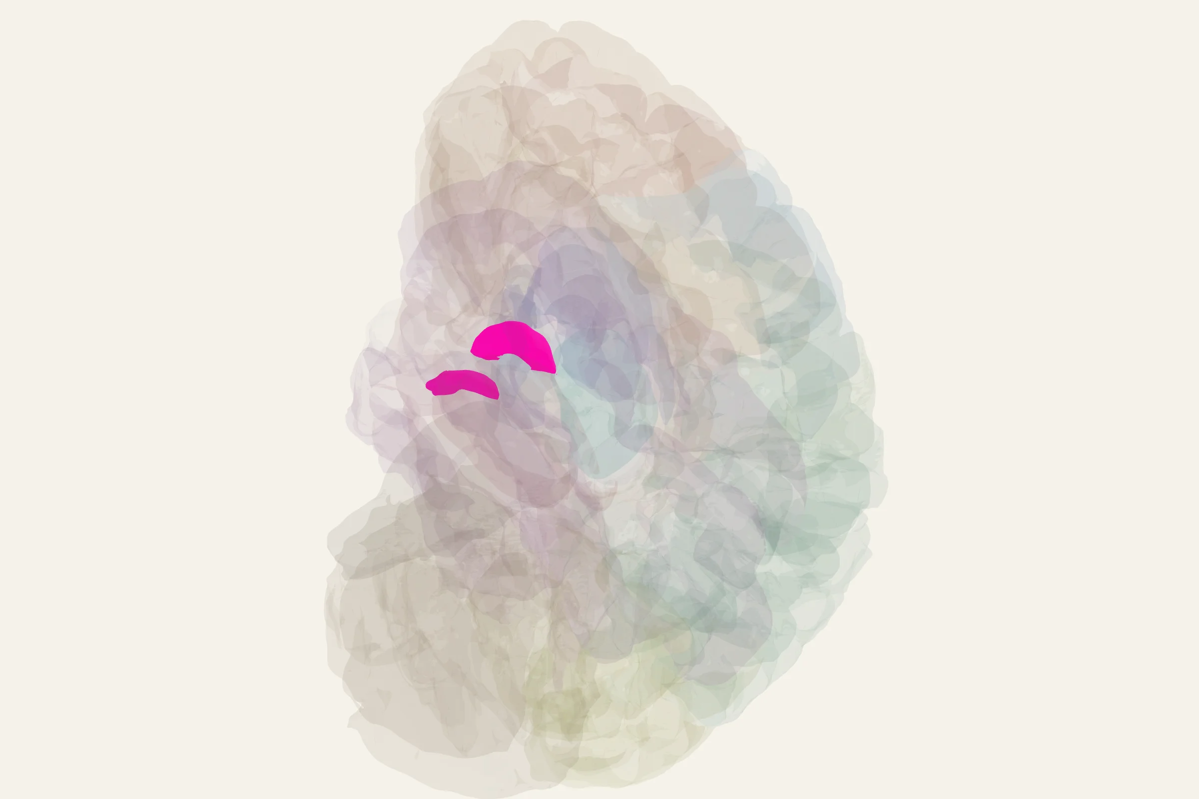
Test this anatomy in the Brain Regions Quiz or open the interactive 3D brain model.
What is the amygdala?
The amygdala is a paired, almond-shaped group of nuclei (the name comes from the Greek amygdale, meaning almond) buried deep in the medial temporal lobe. It is not a single structure but a collection of roughly a dozen nuclei and cortical regions, organized into a handful of functional groups. Together, these nuclei act as the brain's threat-and-salience detector: they evaluate how emotionally significant an incoming stimulus is, then coordinate the body's response through connections to the hypothalamus, brainstem, and cortex. The amygdala is one of the oldest components of the limbic system, and in some contexts it is described as part of the "lizard brain," a colloquial label for the subcortical circuits that handle fast, automatic responses to danger.
Where is the amygdala located?
The amygdala sits in the anterior medial temporal lobe, just rostral to the head of the hippocampus and inferior to the putamen. On the medial surface of the temporal lobe it forms a bulge called the uncus, which is the surface landmark you see first. Roughly two centimeters behind the temporal pole and a few centimeters lateral to the optic chiasm, it rests above the roof of the temporal horn of the lateral ventricle. Because it is hidden beneath the cortical surface, you need a coronal or sagittal cut, or a medial view of the temporal lobe, to see it on a 3D brain model.
What does the amygdala do?
The amygdala's job is to evaluate emotional and behavioral significance and to bias learning, attention, and physiological state toward whatever it judges important. Its specific functions include:
- Detect and learn threats. The amygdala is essential for fear conditioning: pairing a neutral stimulus with an aversive outcome turns the neutral stimulus into a fear cue, and this depends on the basolateral and central nuclei.
- Recognize fearful faces. Damage to the amygdala impairs the ability to read fear, anger, and other negative expressions from facial cues.
- Assign emotional value to sensory input. Visual, auditory, olfactory, and somatosensory signals that reach the amygdala are tagged as pleasant, unpleasant, threatening, or rewarding before being passed on.
- Trigger autonomic and endocrine responses. Through projections to the hypothalamus and brainstem, the amygdala drives the racing heart, dilated pupils, cortisol release, and freezing or flight behavior that accompany fear and stress.
- Modulate emotional memory. Working with the hippocampus, the amygdala strengthens consolidation of memories that carry emotional weight, which is why emotionally charged events are remembered more vividly than neutral ones.
- Drive reward learning and appetitive behavior. The basolateral amygdala contributes to learning which cues predict reward, and it interacts with the nucleus accumbens and ventral striatum in motivated behavior.
- Process social and aggressive behavior. The amygdala contributes to evaluating social signals, dominance, and aggressive responses through its connections to the hypothalamus.
- Shape attention and perception. When the amygdala flags a stimulus as salient, it biases attention and perceptual processing toward that stimulus through feedback to sensory and prefrontal cortices.
Anatomy of the amygdala
The amygdala is conventionally divided into three main nuclear groups, plus a set of small inhibitory clusters that sit between them.
Basolateral nuclear group. This is the largest group and the main input station. It includes the lateral nucleus, basal nucleus, and accessory basal nucleus. These nuclei receive dense projections from sensory cortices, the prefrontal cortex, the entorhinal cortex, and the hippocampus, and they project back to the same areas. The lateral nucleus is the entry point for fear-conditioning information arriving from the thalamus and cortex.
Centromedial nuclear group. Composed of the central nucleus and the medial nucleus, this group is the main output station. The central nucleus projects heavily to the hypothalamus, periaqueductal gray, reticular formation, and other brainstem nuclei, driving autonomic and behavioral fear responses. The medial nucleus is closely linked to olfactory and reproductive behavior circuits.
Cortical (or pale) amygdala. A set of small nuclei near the surface that receive direct input from the olfactory bulb and piriform cortex and pass olfactory information into the rest of the amygdala.
Intercalated cell masses. Small clusters of GABAergic neurons wedged between the basolateral and centromedial groups. They receive input from the prefrontal cortex and inhibit the central nucleus, providing a cortical brake on amygdala output.
Main inputs. The amygdala receives major projections from the medial dorsal and midline nuclei of the thalamus, from unimodal and polymodal sensory cortices, from the prefrontal and anterior cingulate cortices, from the hippocampus and entorhinal cortex, from the insula, and from the olfactory bulb.
Main outputs. Major outputs travel to the hypothalamus, the periaqueductal gray of the midbrain, the reticular formation, the ventral striatum and nucleus accumbens, the mediodorsal thalamus, and back to the prefrontal cortex and hippocampus, forming several closed loops.
What happens when the amygdala is damaged?
Bilateral amygdala damage produces a characteristic set of changes, especially in emotional and social behavior.
- Loss of fear conditioning and reduced fear recognition. The most famous case is the patient known in the literature as S.M., whose bilateral amygdala damage left her unable to experience fear or to recognize fear in facial expressions.
- Klüver-Bucy syndrome. Bilateral damage to the anterior temporal lobes, including the amygdala, produces placidity, hyperorality, hypersexuality, visual agnosia, and a striking loss of fear and aggression.
- Urbach-Wiethe disease. This rare genetic condition causes bilateral calcification that often destroys the amygdala, producing the same kind of fear loss and social flattening.
- Anxiety disorders and PTSD. Functional imaging consistently shows amygdala hyperreactivity in phobias, generalized anxiety disorder, panic disorder, and PTSD, even though the structural integrity of the nucleus is usually intact.
- Temporal lobe epilepsy. The amygdala is one of the most common seizure foci in temporal lobe epilepsy and is often targeted surgically when seizures become drug-resistant.
- Altered social judgment. Damage to the amygdala impairs trust judgments, moral reasoning about intention, and the ability to read negative facial expressions, especially fear and anger.
How to find the amygdala on the 3D brain model
On the 3D brain model at 3d brain model, rotate the brain so you are looking at its ventral or medial surface. The amygdala sits in the anterior medial temporal lobe as a small bulge called the uncus, just in front of the hippocampus and below the basal ganglia. Cutting sagittally through the temporal lobe, or making a coronal section just behind the temporal pole, reveals the amygdala as a rounded gray-matter mass capping the tip of the hippocampal head.
Test yourself
Once you have located the amygdala on the model, quiz yourself on its nuclei, its connections to the hippocampus and prefrontal cortex, and the clinical signs of bilateral damage in the amygdala quiz.
Amygdala key facts
Atlas category: subcortical.
- LeDoux: the 'low road' (thalamus→amygdala) is faster than cortical route by ~12ms
- Amygdala enhances memory for emotional events via norepinephrine in hippocampus
- Fear conditioning: amygdala learns CS-US associations in just one trial
- Fear extinction requires vmPFC inhibiting amygdala — basis of exposure therapy
Amygdala exam tip
Amygdala = fear learning. Two roads: fast (thalamus) and slow (cortex). Extinction = vmPFC inhibiting amygdala, NOT erasing the fear memory.
Common questions
What does the amygdala do?
It supports threat detection, fear conditioning, emotional memory and interpretation of socially important facial signals, especially fear.
How do the amygdala and hippocampus differ?
The amygdala emphasizes emotional significance and can strengthen memory under arousal; the hippocampus binds contextual and episodic details such as what happened, where and when.
Does fear extinction erase an amygdala memory?
No. Extinction is new learning, supported by ventromedial prefrontal inhibition of amygdala responses, rather than deletion of the original fear association.
What part of the brain controls emotions?
Emotion is produced by a circuit, not one structure. The amygdala assigns threat and emotional significance, the insula reads internal bodily state, the anterior cingulate weighs conflict and distress, and prefrontal cortex regulates the response. The amygdala is the part most often meant by the question, but it does not act alone.
Sources
- Purves, Neuroscience, 6th ed., chapters on the limbic system and the emotional brain
- Kandel, Schwartz, and Jessell, Principles of Neural Science, 5th ed., ch. 48 (Emotion)
- Standring, Gray's Anatomy, ch. 23 (Telencephalon)
- StatPearls: Neuroanatomy, Amygdala
- LeDoux, J. The Emotional Brain (background reading on fear-conditioning circuitry)