Hippocampus — Function, Location & Quiz
Updated
The hippocampus is a curved, seahorse-shaped structure buried deep in the medial temporal lobe of the brain, with one hippocampus in each cerebral hemisphere.
The hippocampus forms new declarative memories: it binds the elements of an experience, supports spatial maps and helps consolidate information toward neocortex. It is not a permanent filing cabinet — procedural learning can proceed through other systems.
That separation is the lesson of patient H.M.: after bilateral medial temporal surgery, new episodic learning was devastated but mirror-tracing skill could still improve. Clinically, the hippocampus also matters because it is vulnerable early in Alzheimer’s disease, temporal lobe epilepsy and hypoxic injury.
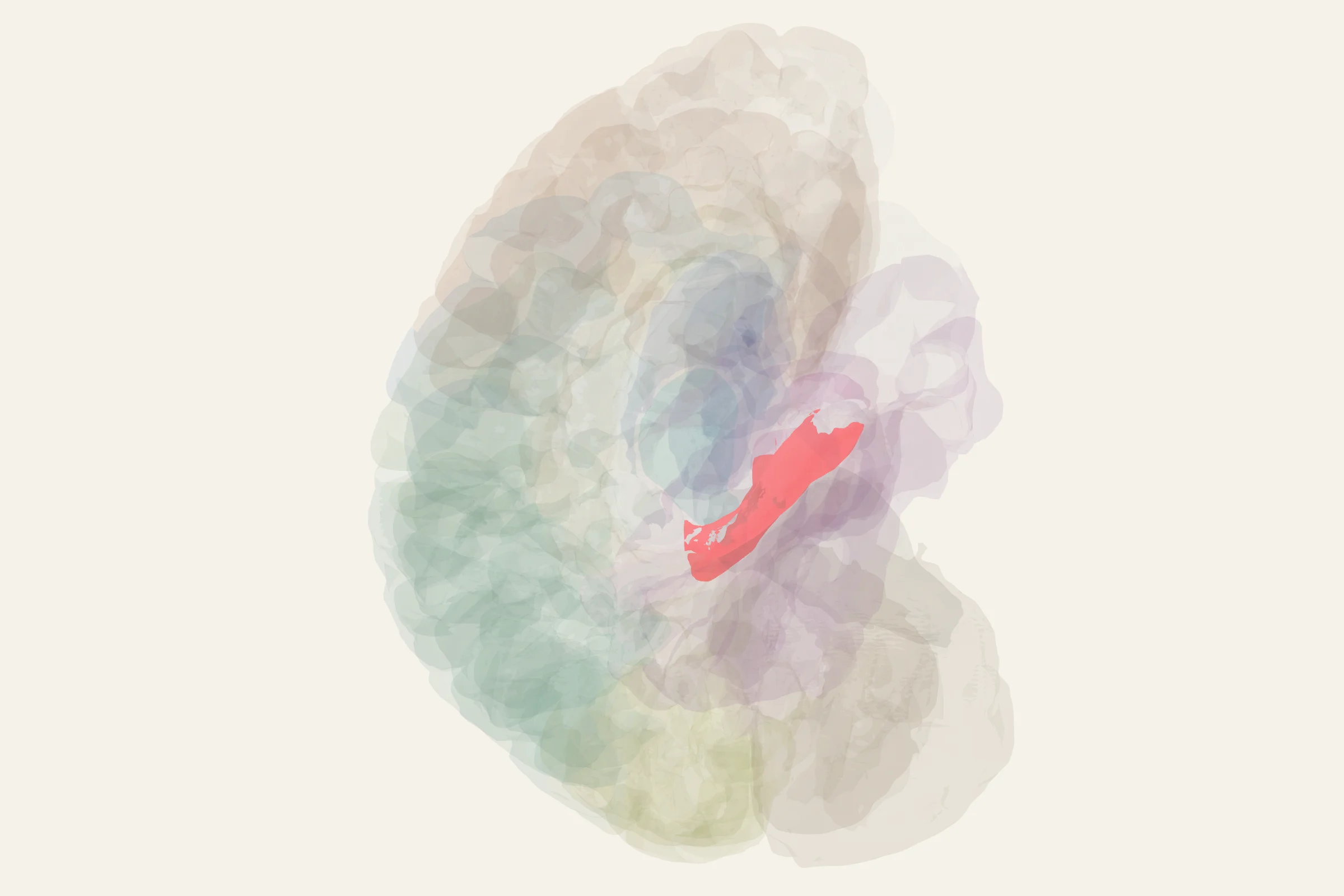
Test this anatomy in the Brain Regions Quiz or open the interactive 3D brain model.
What is the hippocampus?
The hippocampus is a cortical structure that takes its name from the Greek word for "seahorse" because of its curled, C-shaped profile in cross-section. It belongs to the limbic system and is sometimes called the gateway to memory, because without it most new events simply cannot be committed to long-term storage.
You have two hippocampi, one tucked into each medial temporal lobe. Together they are usually described as the hippocampal formation, which also includes the dentate gyrus, the subiculum, and the adjacent entorhinal cortex. When a textbook discusses hippocampus function in psychology or clinical neurology, it is the work of this entire formation that is meant.
The hippocampus in brain networks sits at the meeting point of the cerebral cortex, the diencephalon, and the amygdala. That position is why its output reaches the hypothalamus, the anterior thalamus, and the prefrontal cortex through a single major tract called the fornix.
Where is the hippocampus located?
The hippocampus sits deep in the medial temporal lobe, just behind and below the amygdala, beneath the cortical surface where you would not see it on a superficial brain diagram. It lies along the floor of the temporal horn of the lateral ventricle and curves forward and upward, wrapping around the midbrain.
In practical terms, the hippocampus location in brain anatomy is:
- medial and inferior within the temporal lobe,
- lateral to the midbrain and the thalamus,
- separated from the overlying neocortex by white matter and the parahippocampal gyrus,
- connected to the diencephalon through the fornix.
On a sagittal slice, the structure appears as a curled tube lying almost horizontally inside the temporal lobe. On a coronal slice through the medial temporal lobe, you see the classic interlocking seahorse - the dentate gyrus nested against the cornu ammonis, the curved CA region that gives the hippocampus part of the brain its original name.
What does the hippocampus do?
The brain hippocampus function is best understood as a small set of distinct, experimentally defined jobs. Each one is supported by decades of patient and animal work, and together they explain why hippocampal damage has such a dramatic effect on everyday life.
- Forms new declarative memories. The hippocampus binds together the sights, sounds, smells, timing, and meaning of an event into a single memory that can later be consciously recalled. This is the function of hippocampus most often disrupted by disease.
- Stores spatial maps. Specialized "place cells" in CA1 and CA3 fire whenever an animal or person enters a specific location in its environment, producing an internal cognitive map that supports navigation.
- Consolidates information from short-term to long-term storage. New memories depend on the hippocampus for days to years; over time they become independent of it through a process called systems consolidation, transferring their content to the neocortex.
- Performs pattern separation. The dentate gyrus helps distinguish similar experiences from each other so that overlapping inputs do not become a single confused memory.
- Performs pattern completion. Recurrent circuitry in CA3 can retrieve a complete memory from a partial cue, which is why a smell or song can trigger a vivid episode.
- Provides context for emotional events. Working with the amygdala, the hippocampus tags memories with their time and place, which is the cellular basis of contextual fear learning.
Anatomy of the hippocampus
On any brain diagram hippocampus is usually drawn as a single curled shape, but internally it is divided into sharply defined subfields that each have their own wiring. Understanding these layers is the key to understanding hippocampal anatomy MRI appearance and to interpreting any lesion of this region.
The principal subfields, running from outside in, are:
- Dentate gyrus - a V-shaped granule cell layer that receives the major cortical input through the perforant path from the entorhinal cortex.
- CA3 - large pyramidal cells with extensive recurrent collaterals that connect one CA3 neuron to many others, supporting pattern completion.
- CA1 - the largest pyramidal cell field, the principal output of the hippocampus proper, and the site classically most vulnerable to ischemia.
- Subiculum - the pyramidal cell region between CA1 and the entorhinal cortex, which acts as the major output gateway of the hippocampal formation.
The trisynaptic circuit is the textbook signature pathway and is worth memorizing:
1. Layer II of the entorhinal cortex sends the perforant path to the dentate gyrus.
2. Dentate granule cells send mossy fibers to CA3.
3. CA3 pyramidal cells send Schaffer collaterals to CA1.
4. CA1 and the subiculum return the processed signal to the deep layers of the entorhinal cortex, closing the loop.
The main inputs arrive through the entorhinal cortex, which in turn receives highly processed information from perirhinal cortex, parahippocampal cortex, the amygdala, and prefrontal association areas. Direct inputs also come from the septal nuclei and the supramammillary region of the hypothalamus.
The main outputs leave through the subiculum and travel along the fornix to the mammillary bodies of the hypothalamus, the anterior thalamic nuclei, the septal nuclei, the prefrontal cortex, and the amygdala. Through these projections, hippocampal activity directly modulates the amygdala hippocampus and prefrontal cortex circuit that underlies emotional memory and goal-directed behavior.
What happens when the hippocampus is damaged?
The classic finding is anterograde amnesia, an inability to form new declarative memories. The most famous case is the patient known as H.M., who after bilateral surgical removal of the medial temporal lobes for epilepsy could no longer create new long-term memories of his daily life, even though his working memory and most of his older memories were preserved. His case defined the modern understanding of the brain hippocampus function.
Other well-established clinical associations with hippocampal damage include:
- Alzheimer's disease, in which the hippocampus and entorhinal cortex are among the earliest regions to show atrophy and neurofibrillary tangle pathology, producing the characteristic early loss of recent memory.
- Transient global amnesia, a sudden, temporary loss of anterograde memory usually lasting several hours and almost always triggered by physical or emotional stress.
- Hypoxic-ischemic injury, in which the CA1 subfield is selectively vulnerable, so cardiac arrest, carbon monoxide poisoning, or near-drowning often produces a memory deficit even when other cognition recovers.
- Mesial temporal lobe epilepsy, in which hippocampal sclerosis is a frequent finding and a cause of memory impairment as well as drug-resistant seizures.
- Post-traumatic stress disorder, in which reduced hippocampal volume has been repeatedly observed, although whether the change is a cause, a consequence, or both remains an active research question.
How to find the hippocampus on the 3D brain model
Open the 3D brain model at 3d brain model and rotate the view until you are looking at the medial surface of one temporal lobe; the hippocampus appears as a curled ridge along the floor of the temporal horn of the lateral ventricle, just behind the amygdala and beneath the parahippocampal gyrus. A sagittal cut slightly off the midline, or a coronal cut through the medial temporal lobe, reveals the classic interlocking dentate gyrus and CA fields.
Test yourself
Now that you have worked through hippocampus function, location, and anatomy, head to the quiz section to test yourself on identifying the hippocampal subfields, tracing the trisynaptic circuit, and recognizing the clinical signs of medial temporal lobe damage.
Hippocampus key facts
Atlas category: subcortical. Brodmann areas or landmark: Not a Brodmann area (allocortex, 3-layer).
- Place cells (O'Keefe, Nobel 2014): fire when animal is at a specific location
- Grid cells (Moser & Moser, Nobel 2014): hexagonal spatial firing in entorhinal cortex
- LTP (Long-Term Potentiation) first described in hippocampus (Bliss & Lømo, 1973)
- Adult neurogenesis occurs in dentate gyrus — one of only two known sites
- London taxi drivers (Maguire): larger posterior hippocampi correlating with navigation experience
- Sharp-wave ripples during sleep replay waking sequences (memory consolidation)
Hippocampus exam tip
HM = the most important patient in memory research. Know: anterograde amnesia, preserved procedural memory, intact STM. Trisynaptic circuit: EC → DG → CA3 → CA1.
Common questions
What is the main function of the hippocampus?
It is essential for forming new declarative memories, binding contextual details, spatial navigation and consolidation of memories into distributed cortical networks.
Where is the hippocampus located?
It is a curved medial temporal lobe structure on each side of the brain, connected to cortex chiefly through the entorhinal region.
What did patient H.M. teach us?
His severe anterograde amnesia after bilateral medial temporal resection showed that the hippocampal system is critical for new declarative memory, while preserved mirror tracing demonstrated a separate procedural memory system.
What part of the brain controls memory?
No single part does. The hippocampus is essential for forming new declarative memories, but storage is distributed across neocortex, skills depend on basal ganglia and cerebellum, and emotional memory involves the amygdala. The hippocampus is the part that binds a new experience together; it is not where memories permanently live.
Sources
- Purves, Neuroscience, 6th ed., chapters on the medial temporal lobe and memory systems.
- Kandel, Principles of Neural Science, chapters on the hippocampal formation.
- Squire, Stark, and Clark, "The Medial Temporal Lobe," Annual Review of Neuroscience.
- StatPearls: Neuroanatomy, Hippocampus.
- Nolte, The Human Brain, hippocampal formation section.