Cerebellum — Function, Location & Quiz
Updated
The cerebellum is a hindbrain structure sitting beneath the occipital lobe and posterior to the brainstem. It coordinates voluntary movement, balance, and motor learning, and contributes to certain cognitive and emotional processes.
The cerebellum coordinates movement: it compares intended and actual performance so timing, force and sequence can be corrected, refining action rather than initiating it. Its compact appearance is deceptive because it contains more neurons than the rest of the brain combined, largely in a repeated circuit architecture.
Cerebellar lesions are recognized by coordination errors rather than paralysis: ataxia, dysmetria, intention tremor and impaired rapid alternating movement. The deficits are typically ipsilateral, an exam distinction from the contralateral pattern expected after many cerebral cortical lesions.
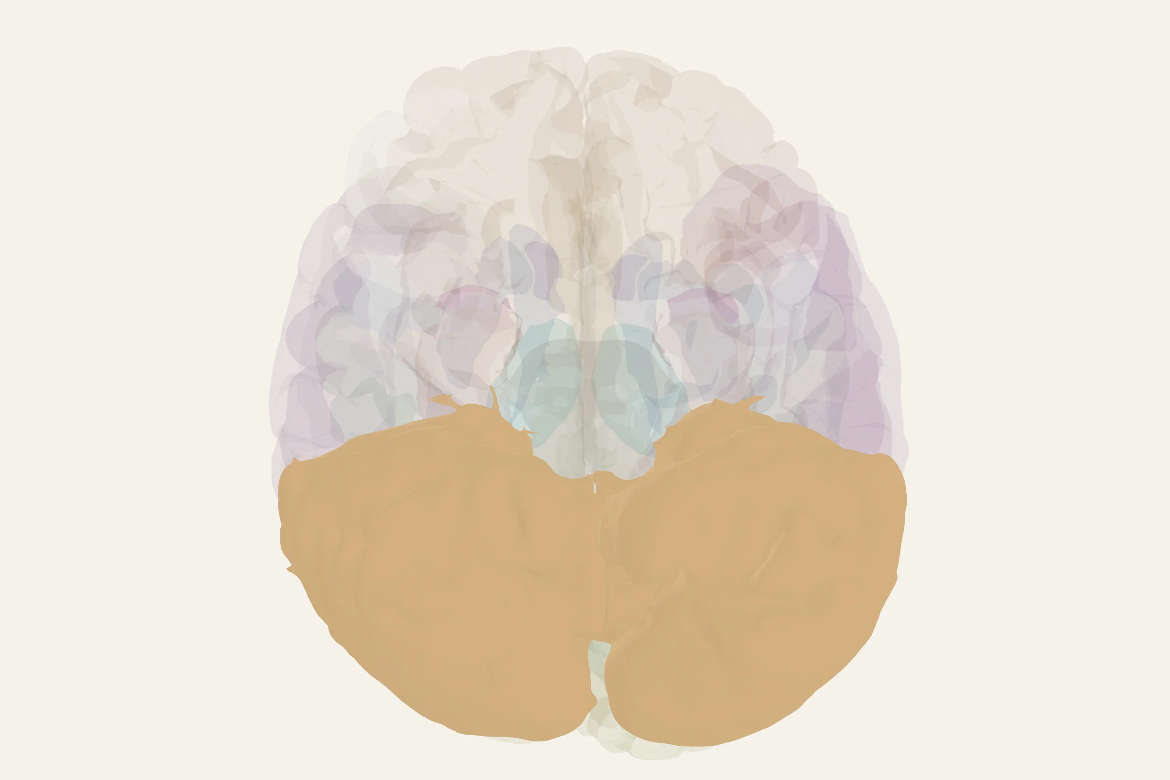
Test this anatomy in the Lesion Localization Quiz or open the interactive 3D brain model.
What is the cerebellum?
The cerebellum (Latin for "little brain") is the largest part of the hindbrain and one of the most distinctive brain parts. It is a paired, cauliflower-shaped mass attached to the back of the brainstem by three thick stalks called peduncles. Its outer surface is a tightly folded sheet of cortex organized into narrow, parallel ridges called folia, which pack a very large surface area into a small space. A midline strip called the vermis separates the two cerebellar hemispheres.
The cerebellum looks like a miniature version of the cerebrum at a glance, but its internal architecture is far more regular and its circuitry more uniform than the cerebral cortex. In humans, the cerebellar hemispheres are unusually large compared with most other mammals, a feature that reflects its growing role beyond motor control.
Where is the cerebellum located?
The cerebellum sits in the posterior cranial fossa, the bony hollow at the back of the skull beneath the tentorium cerebelli. It lies inferior to the occipital lobe of the cerebrum and dorsal (posterior) to the pons and medulla. Those two brainstem structures, together with the cerebellum, make up the hindbrain, or rhombencephalon. The fourth ventricle lies between the cerebellum and the brainstem; cerebrospinal fluid circulates through this space before draining into the central canal of the spinal cord.
On a brain viewed from below, the cerebellum is the rounded, ridged mass you see behind and beneath the brainstem. On a midsagittal section, it appears as the rounded, foliated body tucked under the back of the cerebrum, anchored to the brainstem by the cerebellar peduncles.
What does the cerebellum do?
The cerebellum's classical job is to make movement smooth, accurate, and well-timed. Beyond that, it contributes to several non-motor functions that are now well established in the literature on cerebellum function and cerebellum function psychology:
- Coordinates voluntary movement by comparing the intended movement (signaled by the motor cortex) with the actual movement and correcting errors in real time.
- Maintains balance and posture by integrating vestibular input from the inner ear with proprioceptive signals from the body.
- Times motor sequences, including the precise firing order needed for speech articulation, finger tapping, and rapid alternating movements.
- Drives motor learning, including the adaptation that lets you adjust a saccade, catch a falling object, or ride a bike after the first try.
- Coordinates eye movements, working with brainstem nuclei to keep gaze stable during head motion and to track moving targets.
- Contributes to certain cognitive functions, including aspects of attention, working memory, and language, particularly when the posterior lobe is engaged.
- Modulates emotion and affect, especially through the vermis and flocculonodular lobe; damage here can blunt emotional responsiveness or produce disinhibited behavior.
The cerebellum does not initiate movement; it refines what other motor systems are already doing.
Anatomy of the cerebellum
The cerebellum has three gross anatomical lobes separated by two deep fissures:
- Anterior lobe, separated from the posterior lobe by the primary fissure.
- Posterior lobe, the largest of the three, separated from the flocculonodular lobe by the posterolateral fissure.
- Flocculonodular lobe, the oldest part phylogenetically, also called the vestibulocerebellum because of its close ties to the vestibular system.
Running down the midline is the vermis; flanking it are the two cerebellar hemispheres, which are far larger in humans than in most other mammals. This expansion correlates with the cerebellum's widening role in cognition and language.
The outer cerebellar cortex has three layers, listed from outside in:
- Molecular layer, sparse in cell bodies but rich in parallel fibers and dendritic arbors.
- Purkinje cell layer, a single row of large Purkinje neurons whose elaborate dendrites fan into the molecular layer.
- Granular layer, packed with small granule cells whose axons ascend into the molecular layer.
Beneath the cortex, the white matter (the arbor vitae, or "tree of life") branches into each folium. Embedded within it are four pairs of deep cerebellar nuclei, the only source of output from the cerebellar cortex:
- Dentate nucleus, the largest, involved in planning and coordinating skilled voluntary movement.
- Emboliform and globose nuclei, sometimes grouped as the interposed nuclei, which contribute to limb coordination.
- Fastigial nucleus, the most medial, tied to posture, gait, and the vestibular system.
The cerebellum communicates with the rest of the brain through three peduncles, each a major highway of fibers:
- Superior cerebellar peduncle (brachium conjunctivum), mainly efferent, carrying output from the dentate and interposed nuclei to the red nucleus and thalamus, which then projects to the motor cortex.
- Middle cerebellar peduncle (brachium pontis), purely afferent, carrying input from the contralateral pontine nuclei, which relay information from the cerebral cortex.
- Inferior cerebellar peduncle (restiform body), mostly afferent, carrying spinocerebellar tracts, olivocerebellar fibers from the inferior olive, and vestibular input.
The largest single input to the cerebellum comes from the contralateral inferior olive via climbing fibers, which synapse one-to-one on Purkinje cells. Mossy fibers carry most of the remaining input, including cortical, spinal, and vestibular information.
What happens when the cerebellum is damaged?
Cerebellar damage produces a recognizable cluster of signs, classically grouped as the cerebellar syndrome:
- Ataxia, a wide-based, unsteady gait with poor balance.
- Dysmetria, the inability to judge distance, so a finger overshoots or undershoots a target on the finger-to-nose test.
- Intention tremor, a tremor that worsens as the hand approaches its target.
- Dysdiadochokinesia, difficulty performing rapid alternating movements such as pronation and supination.
- Nystagmus, rhythmic eye movements that drift and snap back.
- Scanning or staccato speech, in which syllables are broken apart.
- Hypotonia, reduced muscle tone on the affected side.
Damage to the vermis, as in chronic alcoholism, produces truncal ataxia and gait instability even when limb coordination is normal. Damage to the flocculonodular lobe or vestibulocerebellum disrupts balance and eye movements most prominently.
When injury extends beyond the motor cerebellum, particularly into the posterior lobe and vermis, the result can be cerebellar cognitive affective syndrome, characterized by executive dysfunction, impaired visuospatial reasoning, blunted affect, and language irregularities. This syndrome was described by Jeremy Schmahmann and colleagues and is now widely accepted.
Common causes of cerebellar injury include stroke, tumors, multiple sclerosis, paraneoplastic degeneration, alcohol-related cerebellar degeneration, and inherited ataxias such as Friedreich's ataxia and the spinocerebellar ataxias.
How to find the cerebellum on the 3D brain model
Open the viewer at 3d brain model and rotate the brain so you are looking at it from behind and slightly below. The cerebellum is the rounded, ridged mass tucked under the occipital cortex and sitting against the brainstem. A midsagittal cut through the center of the brain reveals its full "tree of life" branching and its attachment to the pons and medulla.
Test yourself
Now that you have the cerebellum's parts and functions in mind, head to the cerebellum quiz on brainquiz.study to label its lobes, deep nuclei, and peduncles on a rotating 3D brain. You can drill by region, name the structure, or flip into function mode to match each area to what it does.
Cerebellum key facts
Atlas category: cerebellum.
- Contains MORE neurons than the rest of the brain combined (~69 billion granule cells)
- Marr-Albus theory: climbing fibers teach cerebellar cortex via LTD (long-term depression)
- Internal models: cerebellum predicts sensory consequences of movement (forward model)
- Eyeblink conditioning — cerebellum is necessary and sufficient (Thompson)
Cerebellum exam tip
Cerebellum = ipsilateral deficits (unlike cerebral cortex which is contralateral). IPSILATERAL ataxia, dysmetria, intention tremor.
Common questions
What does the cerebellum do?
It fine-tunes movement timing and accuracy, supports balance and posture, learns from motor error and also contributes to cognition and affect.
Why are cerebellar deficits ipsilateral?
Cerebellar motor pathways effectively cross twice before influencing the body, so a lesion generally disrupts coordination on the same side.
What are classic signs of a cerebellar lesion?
Ataxia, dysmetria, intention tremor and dysdiadochokinesia are high-yield signs. Weakness alone is not the defining deficit.
What part of the brain controls balance?
The cerebellum, working with the vestibular system. Vestibular input from the inner ear reaches brainstem vestibular nuclei, and the cerebellum uses it to correct posture and gait moment by moment. Damage produces unsteadiness and a wide-based walk rather than weakness.
Sources
- Purves, Neuroscience, 6th ed., ch. 21 on the cerebellum
- Kandel, Principles of Neural Science, 5th ed., ch. 42
- Standring, Gray's Anatomy for Students, cerebellar chapter
- StatPearls: Neuroanatomy, Cerebellum
- Schmahmann, "The cerebellum and cognition," 1991, and related work on cerebellar cognitive affective syndrome