Brainstem — Function, Location & Quiz
Updated
The brainstem is the stalk-like lower part of the brain that connects the cerebrum, diencephalon, and cerebellum to the spinal cord.
The brainstem is both a conduit and a life-support structure: long motor and sensory tracts pass through it while local nuclei regulate breathing, circulation, arousal and most cranial nerves. Treating it as one undifferentiated stalk hides the practical map of midbrain, pons and medulla.
Small lesions can therefore combine crossed sensory or motor findings with a cranial-nerve deficit at the level of injury. Ventral pontine damage can produce locked-in syndrome while awareness survives, showing why loss of movement is not the same as loss of consciousness.
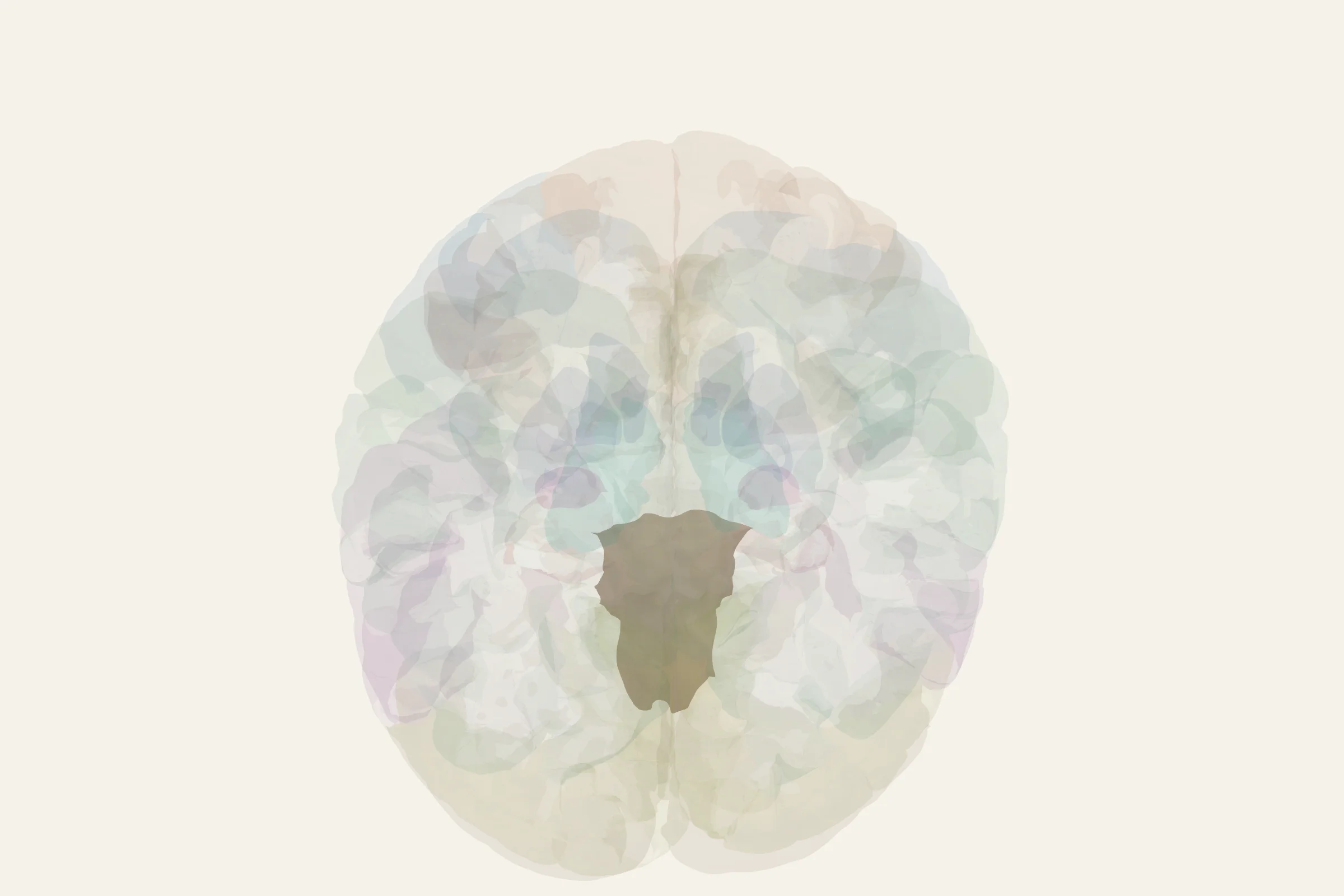
Test this anatomy in the Lesion Localization Quiz or open the interactive 3D brain model.
What is the brainstem?
The brainstem is the most caudal portion of the brain, the segment that sits between the diencephalon above and the spinal cord below. Anatomists group it into three continuous structures: the midbrain (mesencephalon), the pons, and the medulla oblongata. Together these regions house the cranial nerve nuclei for nerves III through XII, the long ascending and descending tracts that link the forebrain to the spinal cord, and a dense core of neurons called the reticular formation. The brainstem is sometimes described as the oldest part of the brain from an evolutionary standpoint, because the same basic circuitry supports breathing, arousal, and sensorimotor relay in every vertebrate. Despite being only a few centimeters long, it carries an outsized share of the central nervous system's traffic and is essential to survival.
Where is the brainstem located?
The brainstem occupies the midline at the base of the skull, sitting in the posterior cranial fossa. Superiorly, it emerges from the underside of the diencephalon, just below the thalamus and hypothalamus. Posteriorly, it lies against the cerebellum, connected to it by the three cerebellar peduncles. Inferiorly, it narrows into the spinal cord at the level of the foramen magnum. From a ventral view, you can see the cerebral peduncles of the midbrain, the bulging belly of the pons, and the medullary pyramids flanking the midline. On a midsagittal cut, the brainstem appears as a roughly vertical column about 7–8 cm long in adults, with the cerebellum tucked behind it and the pituitary suspended from the hypothalamus in front.
What does the brainstem do?
The brainstem performs five overlapping jobs that bridge the body and the rest of the brain.
- Controls breathing. The medullary respiratory center (dorsal and ventral respiratory groups) and the pontine respiratory group set the rhythm and depth of ventilation in response to CO₂, O₂, and pH levels in the blood.
- Controls heart rate and blood pressure. The cardiovascular center in the medulla adjusts cardiac output and vessel tone through sympathetic and parasympathetic outflow, which is why brainstem injury often produces rapid circulatory collapse.
- Maintains arousal and consciousness. The ascending reticular activating system in the core of the brainstem sends diffuse projections to the thalamus and cortex; damage to this system is a common cause of coma.
- Relays signals between the brain and spinal cord. Nearly every major tract — corticospinal, corticobulbar, medial lemniscus, spinothalamic — passes through the brainstem, and many decussate here.
- Coordinates reflexes. Sneezing, coughing, gagging, swallowing, and vomiting are organized by brainstem nuclei and circuitry that link sensory input to motor output without cortical involvement.
Anatomy of the brainstem
Midbrain (mesencephalon)
The midbrain is the most rostral segment, sitting just below the thalamus. Its dorsal surface, the tectum, carries the superior colliculi (visual reflexes and saccadic eye movements) and the inferior colliculi (auditory relay to the medial geniculate). Its ventral surface shows the two cerebral peduncles, which carry the descending corticospinal and corticobulbar fibers. Inside, the tegmentum contains the red nucleus, the substantia nigra (whose dopamine-containing neurons degenerate in Parkinson's disease), cranial nerve nuclei III and IV, and the periaqueductal gray, a region involved in pain modulation and autonomic control.
Pons
The pons is the bulging middle segment of the brainstem, easily recognized by its ventral swelling. Its dorsal portion, the pontine tegmentum, houses cranial nerve nuclei V, VI, VII, and VIII, the locus coeruleus, and parts of the reticular formation. Its ventral basis pontis contains the pontine nuclei, which receive input from the cerebral cortex and project across the midline into the contralateral cerebellum through the middle cerebellar peduncle, making the pons the main relay station between cerebrum and cerebellum.
Medulla oblongata
The medulla is the most caudal segment of the brain, continuous with the spinal cord below the foramen magnum. On its ventral surface, the medullary pyramids carry the corticospinal tract, and most of those fibers decussate here at the pyramidal decussation. The medulla also contains the inferior olivary nucleus (a major source of climbing-fiber input to the cerebellum), the gracile and cuneate nuclei (relays for fine touch, vibration, and conscious proprioception), and the nuclei of cranial nerves IX, X, XI, and XII. Inside the reticular core, scattered cholinergic and noradrenergic cell groups contribute to arousal and autonomic regulation.
Blood supply
The brainstem receives blood almost entirely from the posterior circulation. The posterior cerebral arteries supply most of the midbrain. Pontine branches of the basilar artery feed the pons. The medulla is supplied by the anterior spinal artery and the posterior inferior cerebellar artery. Because each brainstem artery feeds a defined column of nuclei and tracts, vascular occlusions produce the classic brainstem syndromes described below.
Inputs and outputs
- Afferent inputs come from the cerebral cortex (corticobulbar and corticospinal fibers), the cerebellum (cerebellothalamic feedback), the spinal cord (spinothalamic and spinoreticular tracts), sensory ganglia of the cranial nerves, and special senses including vestibular, auditory, and taste pathways.
- Efferent outputs leave through cranial nerves III–XII, through the corticospinal and corticobulbar fibers descending from the cortex, and through reticulospinal, vestibulospinal, and tectospinal tracts descending into the spinal cord to influence posture, gaze, and autonomic tone.
Cranial nerves from the brainstem
Ten of the twelve cranial nerves have their nuclei inside the brainstem.
- Midbrain: III (oculomotor), IV (trochlear)
- Pons: V (trigeminal), VI (abducens), VII (facial), VIII (vestibulocochlear)
- Medulla: IX (glossopharyngeal), X (vagus), XI (accessory), XII (hypoglossal)
The spinal accessory nerve (XI) is a partial exception: its cranial root exits from the medulla, but its spinal rootlets emerge from the upper cervical cord and join the brainstem outflow before exiting the jugular foramen. Only the olfactory (I) and optic (II) nerves arise outside the brainstem, directly from the forebrain.
What happens when the brainstem is damaged?
Brainstem damage is dangerous because the same small region that regulates vital functions also carries the long tracts that control movement and sensation. Common causes include posterior-circulation stroke, traumatic brain injury, multiple sclerosis plaques, brainstem gliomas, and compression from basilar artery aneurysms. A few named syndromes illustrate the consequences.
- Weber syndrome — a midbase midbrain stroke affecting the cerebral peduncle and the exiting oculomotor nerve, producing contralateral hemiparesis with ipsilateral ptosis and a "down and out" eye.
- Wallenberg syndrome (lateral medullary syndrome) — usually from occlusion of the vertebral or posterior inferior cerebellar artery; causes ipsilateral loss of facial pain and temperature, contralateral loss of body pain and temperature, hoarseness, vertigo, dysphagia, and Horner's syndrome.
- Medial medullary syndrome — infarction of the anterior spinal artery territory; produces contralateral hemiparesis, contralateral loss of position and vibration sense, and ipsilateral tongue deviation.
- Locked-in syndrome — a ventral pontine lesion that interrupts corticospinal and corticobulbar tracts, leaving the patient awake and aware but unable to move or speak except for vertical eye movements and blinking.
- Central sleep apnea (Ondine's curse) — failure of the automatic breathing drive after bilateral medullary damage, requiring ventilator dependence especially during sleep.
- Brain death — clinically defined by the irreversible loss of all brainstem reflexes (pupillary, corneal, gag, cough, caloric) together with coma and apnea.
Because the cranial nerve nuclei sit here, brainstem lesions also produce focal signs such as diplopia, dysarthria, dysphagia, tongue deviation, and altered facial sensation that help localize the level of injury.
How to find the brainstem on the 3D brain model
Open the 3D brain model at 3d brain model and rotate to a midsagittal or ventral view. The brainstem appears as a vertical column in the center, with the cerebellum sitting behind it and the thalamus directly above. Selecting the cut-plane tool along the midline cleanly exposes the midbrain, pons, and medulla in sequence, and highlights the superior colliculus, substantia nigra, medullary pyramids, inferior olive, and facial nucleus as named subregions.
Test yourself
Use the brain stem diagram quiz and the labeled brain stem practice on the structure tab to identify the midbrain, pons, and medulla, and to match each cranial nerve with the brainstem segment where its nucleus lies. Your quiz results feed into a personalized review list, so each session targets the structures you missed.
Brainstem key facts
Atlas category: brainstem.
- Reticular activating system: damage → coma (consciousness requires brainstem)
- Locus coeruleus (pons) = norepinephrine source → arousal, attention
- Raphe nuclei (midline) = serotonin source → mood, sleep
- Substantia nigra (midbrain) = dopamine source → movement (Parkinson's)
- VTA (midbrain) = dopamine source → reward (mesolimbic pathway)
Brainstem exam tip
Brainstem = 3 parts: midbrain (superior), pons (middle), medulla (inferior). Know the neurotransmitter sources: LC=NE, Raphe=5HT, SNc=DA, VTA=DA.
Common questions
What are the three parts of the brainstem?
From superior to inferior they are the midbrain, pons and medulla. The medulla continues into the spinal cord.
What does the brainstem control?
It regulates breathing, heart rate, blood pressure, arousal and pain modulation, contains cranial-nerve nuclei, and carries major ascending and descending pathways.
What is locked-in syndrome?
A ventral pontine lesion can interrupt descending motor pathways and leave a person conscious but unable to move most of the body, often preserving vertical eye movement for communication.
What part of the brain controls breathing?
The brainstem, specifically respiratory centres in the medulla and pons. The medulla sets the basic rhythm and pontine centres shape its timing, all running without conscious effort. This is why brainstem injury is life-threatening in a way that damage to most cortical regions is not.
Sources
- Purves, D., et al. (eds.), Neuroscience, 6th ed., Sinauer/Oxford University Press, chapters 1, 18, and 26
- Standring, S. (ed.), Gray's Anatomy, 42nd ed., Elsevier, sections on brainstem and cranial nerves
- Haines, D. E., Neuroanatomy Atlas in Clinical Context, 10th ed., Wolters Kluwer
- StatPearls: Neuroanatomy, Brainstem (Treuting et al.)
- Brazis, P. W., Localization in Clinical Neurology, 7th ed., chapter on brainstem lesions