Occipital Cortex — Function, Location & Quiz
Extrastriate occipital cortex is what happens after V1 has registered the basic visual layout. Parallel areas develop different emphases—V4 for colour and form, V5/MT for motion—before information continues toward temporal and parietal association systems.
Selective lesions show that seeing is not one indivisible ability. Achromatopsia removes cortical colour experience, while akinetopsia makes movement appear as disconnected positions; neither is explained by ordinary eye disease.
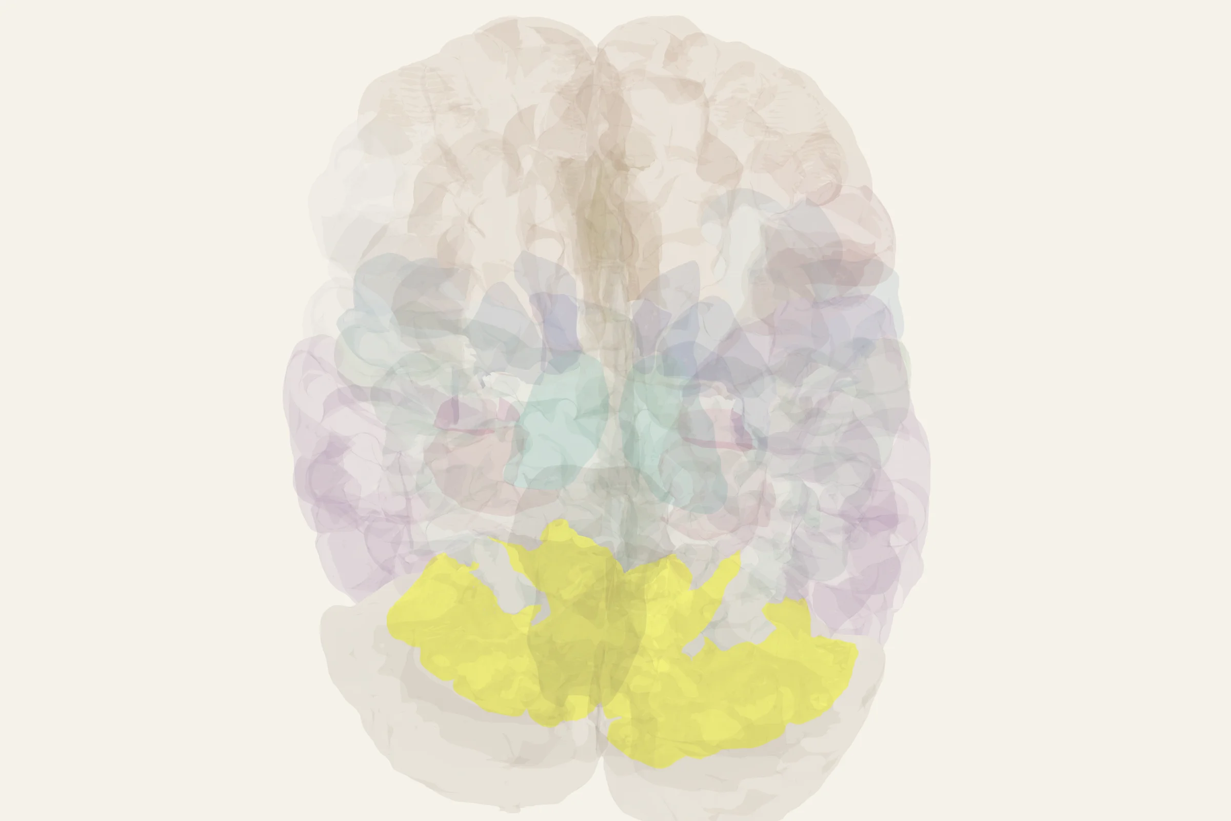
Test this anatomy in the Visual Field Defect Quiz or open the interactive 3D brain model.
Where is the Occipital Cortex located?
Visual association, motion processing, color processing.
What does the Occipital Cortex do?
- Higher-order visual processing beyond V1
- V4: color perception and shape processing
- V5/MT: motion perception and optic flow
- V3: dynamic form processing
Occipital Cortex pathways and connections
- V1 → V2 → V3/V4/V5 (parallel processing streams)
- V5/MT → MST (optic flow for self-motion)
- V4 → IT cortex (color + form for object recognition)
What happens when the Occipital Cortex is damaged?
- V4 lesion → achromatopsia (cortical color blindness)
- V5/MT lesion → akinetopsia (motion blindness — patient LM)
- Patient LM (Zihl): couldn't see motion, tea appeared to 'jump' in cup
Occipital Cortex key facts
Atlas category: cortical. Brodmann areas or landmark: BA 18, 19 (V2, V3, V4, V5/MT — extrastriate).
- Zeki: V4 is the 'color center,' V5/MT is the 'motion center'
- V5/MT neurons have large receptive fields and respond to coherent motion
- TMS to V5/MT temporarily impairs motion direction judgments
Occipital Cortex exam tip
Extrastriate = everything after V1. V4 = color. V5/MT = motion. Know Patient LM for motion blindness!
Common questions
How is occipital association cortex different from V1?
V1 is the first cortical visual stage. Extrastriate areas in Brodmann areas 18 and 19 perform higher-order analyses such as colour, form, motion and optic flow.
Which visual area processes motion?
Area V5, also called MT, is strongly specialized for motion direction and coherent movement. Damage can cause akinetopsia, or cortical motion blindness.
What happens after a V4 lesion?
A V4 lesion can produce cerebral achromatopsia, in which colour perception is lost or severely impaired despite functioning eyes.